Deep-sea chemosynthetic ecosystems represent a frontier window into the evolution of life on Earth. In lightless, high-pressure, strongly reducing cold seep and hydrothermal vent environments, chemosymbiotic systems support thriving benthic biological communities. Previously known large chemosymbiotic animals (such as deep-sea mussels, tube worms, white clams, and lucinid bivalves) mainly accommodate symbiotic bacteria through esophageal glands or within single-membrane vacuoles in bacteriocytes. However, whether hosts could evolve dedicated independent "organelles" specifically serving chemosymbiosis had never been reported.
Recently, Professor Qian Peiyuan from the Guangzhou Marine Laboratory, together with Professor Sun Jin from the Ocean University of China, Professor Qiu Jianwen from Hong Kong Baptist University, and multiple domestic and international research teams, published a research paper online in the international comprehensive academic journal Nature Communications, entitled "A multilamellar organelle for chemosymbiosis in an aplacophoran mollusc adapted to anoxic cold seep sediment". From organ, cellular to molecular scales, using cutting-edge technologies including three-dimensional structural reconstruction, ultrastructural electron microscopy analysis, whole-genome sequencing, spatial transcriptomics, and spatial metabolomics, the study discovered a completely novel host-bacteria symbiotic structure in the deep-sea aplacophoran mollusk Chaetoderma shenloong from the South China Sea cold seep, and identified a specialized multilamellar organelle dedicated to housing and cultivating sulfur-oxidizing symbiotic bacteria — "dracosphera" (meaning "dragon sphere"), revealing the adaptation mechanism of deep-sea life to extreme hypoxia. This research is an important scientific achievement under the framework of CliMetS and MOCSI, the UN Decade programmes, focusing on scientific research, international cooperation, and collaborative observation of global cold seeps and related interfaces, bringing together research forces from multiple countries and regions, and committed to advancing research on deep-sea cold seep ecosystems, methane cycling, and their response mechanisms to environmental changes.
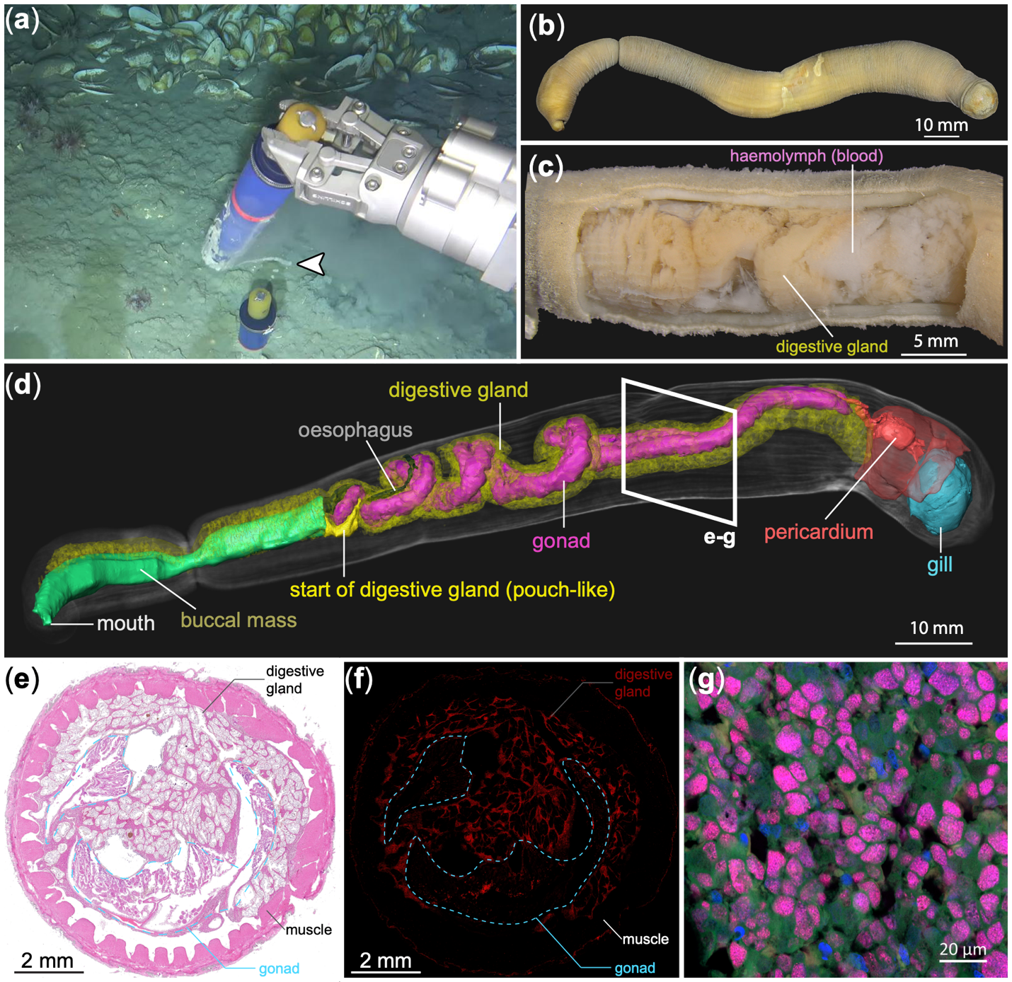
Figure 1. Deep-sea cold seep habitat, external morphology, and internal symbiotic organs of Chaetoderma shenloong. Three-dimensional anatomical reconstruction and fluorescence in situ hybridization show that its significantly enlarged reticulated digestive gland accommodates abundant symbiotic bacteria.
Exploring the Haima Cold Seep of the South China Sea: The "Deep-Sea Dragon" Buried in Deep Anoxic Mud Layers
Chaetoderma shenloong belongs to the class Caudofoveata within the phylum Mollusca, and is the first known vermiform aplacophoran mollusk associated with deep-sea chemosynthetic habitats. Typical caudofoveatan species usually measure only about 10 mm in body length and mostly live in shallow surface sediment layers, whereas C. shenloong can reach over 150 mm in body length, exhibiting a remarkable "deep-sea gigantism" trait. Owing to its unique evolutionary status and morphological characteristics, this species was selected by the World Register of Marine Species (WoRMS) as one of the "Top 10 Marine New Species of 2024" worldwide. What is surprising is its extreme habitat: the research team discovered during sampling at the "Haima" cold seep area in the South China Sea that these animals are densely buried tens of centimeters deep in the black reducing mud layer below the sediment surface; in situ microelectrode measurements revealed that the sediment in this area becomes completely anoxic just 3 mm below the surface, and deeper layers are enriched with high concentrations of hydrogen sulfide.
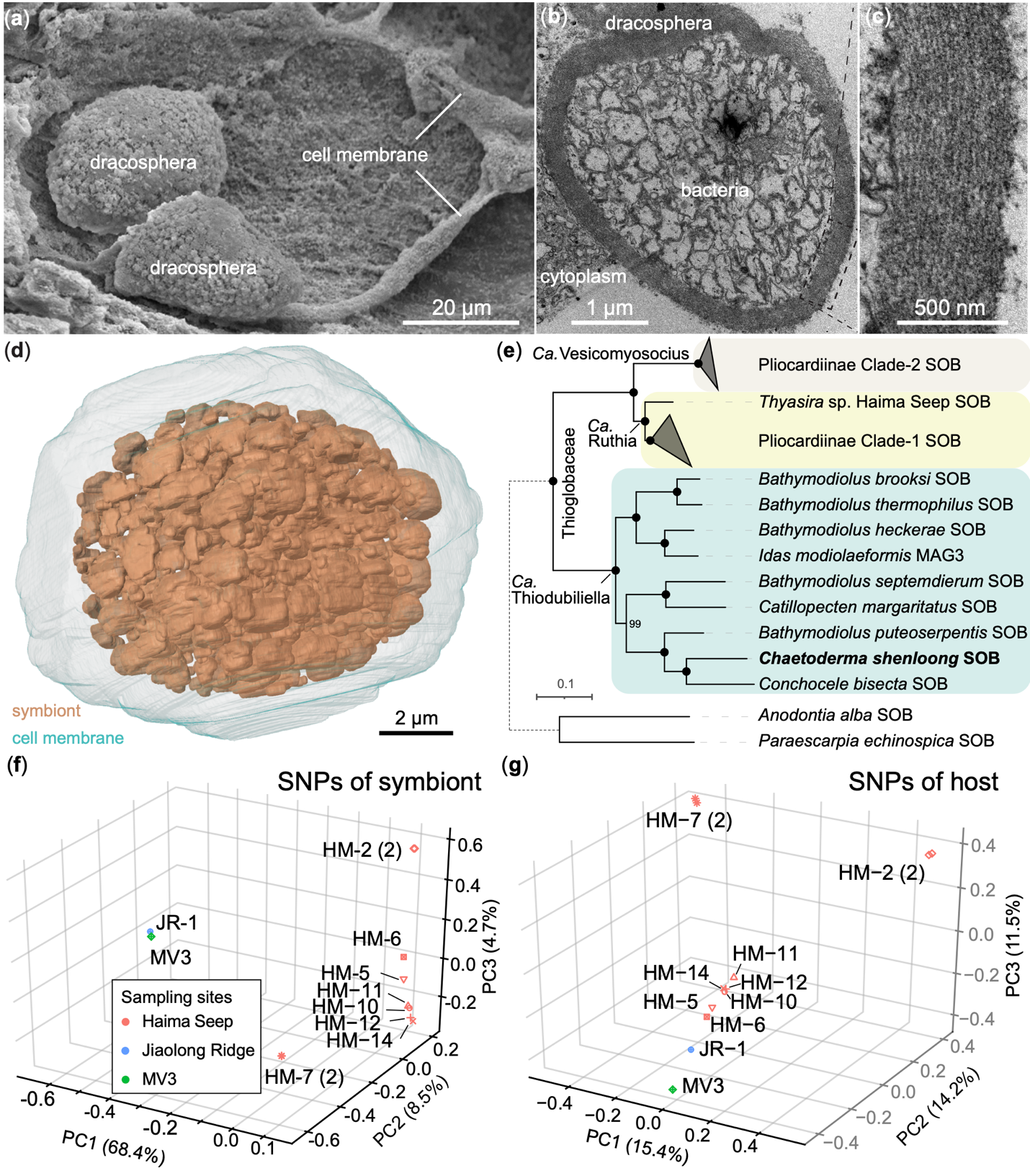
Figure 2. Electron micrographs and three-dimensional reconstruction of the dracosphera. Sulfur-oxidizing symbiotic bacteria are densely distributed inside the spherical structure, wrapped by multiple membrane layers.
Digestive Tract Degeneration and Discovery of the Specialized Multilamellar Organelle "Dracosphera"
Anatomical studies and micro-CT (µ-CT) three-dimensional reconstruction revealed that the digestive system of C. shenloong has undergone dramatic specialization and degeneration during evolution. Its radula is highly degenerate, and the midgut, hindgut, and anus posterior to the esophagus are almost completely absent. Instead, the digestive gland is highly enlarged, distributed in a complex reticulated and diffuse pattern, and occupies most of the body cavity. Fluorescence in situ hybridization (FISH) and carbon-nitrogen stable isotope analysis confirmed that this specialized digestive gland is the core symbiotic organ where chemosynthesis takes place, densely enriched with abundant symbiotic bacteria signals.
Further combining transmission electron microscopy (TEM) with high-precision focused ion beam scanning electron microscopy (FIB-SEM) three-dimensional analysis, the researchers revealed its unprecedented subcellular ultrastructure — completely different from previously known chemosymbiosis where bacteria only colonize the host epithelial surface or single-membrane vacuoles. Within the bacteriocytes of the digestive gland, spherical vesicles with diameters of approximately 10 to 15 micrometers are densely distributed, each structure wrapped by as many as 20 or more layers of dense lipid membranes, with an average membrane wall thickness of 506 nanometers. The research team named this novel organelle "dracosphera" (dragon sphere), combining the Latin words draco (dragon) and sphera (sphere). Each dracosphera tightly encloses hundreds to thousands of sulfur-oxidizing symbiotic bacteria (Candidatus Thiodubiliella), with an average of up to 1,629 symbiotic bacteria accommodated in a single host cell. Although the volume of the "dracosphera" is comparable to that of a typical eukaryotic cell, the interior of the dracosphera completely lacks a eukaryotic nucleus and lysosomes, thus ruling out the possibility that it functions as a conventional lysosome for digestion, indicating that the host has developed a sophisticated maintenance mechanism to provide long-term shelter and cultivation for the symbiotic bacteria.
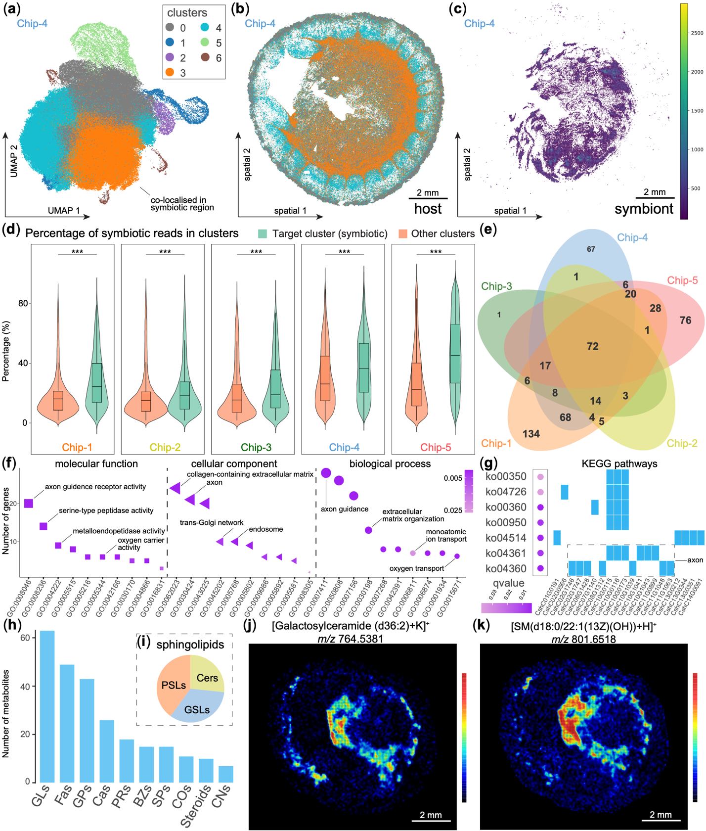
Figure 3. Spatial transcriptomic and spatial metabolomic analyses reveal gene expression and lipid distribution characteristics in symbiotic tissues, providing molecular mechanisms for studying the composition and maintenance of dracosphera.
Combined Spatial Multi-Omics: Revealing the "Oxygen Barrier" and Nutrient Exchange Strategies Under Extreme Hypoxia
To systematically elucidate the unique physiological and metabolic functions of the "dracosphera," the team constructed a high-precision genome of this symbiotic system (including the host chromosome-level genome and the circular symbiont genome), and conducted multi-dimensional analysis integrating metatranscriptomics, in situ spatial transcriptomics, and desorption electrospray ionization mass spectrometry imaging (DESI-MSI) spatial metabolomics. Genomic analysis showed that the symbiont genome has been highly reduced to 1.14 Mb, possesses microaerophilic and facultative anaerobic metabolic potential, is highly dependent on the denitrification pathway as an electron acceptor, and specifically retains the cbb3-type cytochrome c oxidase with extremely high affinity under ultra-low oxygen conditions. Since the host needs to dive deep into low-oxygen mud layers to absorb reduced sulfide, the internal oxygen supply is extremely scarce; the tight multilamellar physical configuration of the "dracosphera," similar to the myelin sheath of mammalian neuronal axons or vesicles of nitrogen-fixing bacteria, can play a key role as an oxygen diffusion barrier, blocking oxygen from contacting the symbiotic bacteria, thereby prioritizing the precious trace oxygen supply to host mitochondria for energy metabolism, achieving optimal energy efficiency in extremely hypoxic habitats.
Meanwhile, spatial metabolomics and spatial transcriptomics revealed the neuron-like differentiation characteristics and material flow direction of this multilamellar membrane system. Spatial metabolomics showed that the symbiotic digestive gland is significantly enriched with 15 sphingolipid molecules including galactosylceramide (GalCer) and sphingomyelin (SM); Raman spectroscopy also confirmed that dracospherae are in a high-lipid state. Spatial transcriptomics detected high-level coordinated expression of genes related to neuronal axon development (such as fasciclin 2, semaphorins) as well as multiple oxygen-transporting hemocyanins, demonstrating the molecular basis for the host to actively construct and dynamically regulate this multilamellar membrane interface. Furthermore, a large number of lysosomes are densely distributed in the host cytoplasm outside the dracosphera, and hydrolase genes such as serine peptidases and metallopeptidases are significantly highly expressed in the symbiotic region, suggesting that the host may periodically regulate local disintegration of the multilamellar membrane to controllably release symbiotic bacteria into the cytoplasm for lysosomal enzymatic digestion, thereby absorbing essential amino acids and organic nutrients that the host cannot synthesize itself, completing efficient "grazing-style" nutrient conversion.
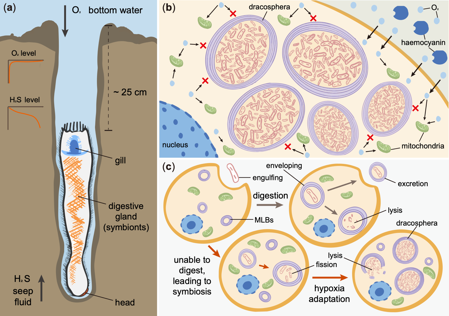
Figure 4. Schematic diagram of the function and evolution of Chaetoderma shenloong and dracosphera. The research team proposes that the multilamellar membrane may restrict oxygen diffusion to symbiotic bacteria and may have evolved from ancestral multilamellar structures used for processing bacteria. The mechanisms shown in the figure are research hypotheses.
Scientific Implications and Global Ecological Significance
Throughout the long evolutionary history of eukaryotes, the integration and transformation of exogenous symbiotic bacteria into organelles (such as mitochondria, chloroplasts, and the recently confirmed nitrogen-fixing nitroplast in algae) has been a decisive event driving major leaps in life. The discovery of the "dracosphera" demonstrates a completely new pathway by which multicellular animals have evolved specialized organelles to harness chemosymbiotic bacteria, providing a new case for exploring eukaryotic organellogenesis and endosymbiotic evolution.
In addition, researchers have also detected C. shenloong in the mud volcano cold seep off Kyushu, Japan, and in the F cold seep sediments of the northern South China Sea. Combined with the known giant caudofoveatan relative species from the Gulf of Mexico reaching 40 cm in body length, this indicates that large chemosymbiotic aplacophorans relying on "dracospherae" to adapt to deep anoxic environments may be widely distributed in global deep-sea cold seeps. Buried beneath the seafloor surface, they carry out efficient dark carbon fixation and hydrogen sulfide consumption, making them long-underestimated important contributors to deep-sea biogeochemical cycling.
The co-corresponding authors of the paper are Professor Sun Jin, Professor Qian Peiyuan, and Professor Qiu Jianwen; the co-first authors include Dr. Li Yunlong and Senior Researcher Chen Chong. This work was supported by the National Key Research and Development Program, Laoshan Laboratory, Shandong Provincial Natural Science Foundation, Guangzhou Marine Laboratory, the Jockey Club Centre for Climate and Sustainability, and the Hong Kong Research Grants Council, among other sources.
Attachment download:
